glvd-great-lakes-veterinary-dermatology-banner (1).png
great-lakes-veterinary-dermatology (1).webp

Great Lakes Veterinary Dermatology

Brookfield, WI · Board-certified Dermatology

Great Lakes Veterinary Dermatology specializes in the treatment and management of all skin disorders affecting dogs and cats, including allergy treatment, immune-mediated disorders, and ear and skin infections.

Learn moreRequest an appointment

Services at This Location

Hyperbaric Oxygen Therapy

Wound healing is a subject of great interest and involvement for the surgeon. While much of the physiology of wound healing is understood, gaps still exist in our understanding of the phenomenon. The surgeon attempts to modify the wound milieu by various means at his disposal. One such method is hyperbaric oxygen therapy (HBOT).HBOT is the use of 100% oxygen at pressures greater than atmospheric pressure. The patient breathes 100% oxygen intermittently while the pressure of the treatment chamber is increased to greater than 1 atmosphere absolute (ATA).[] HBOT has had very exciting and interesting beginnings. In 1620, Drebbel developed a one-atmosphere diving bell.[] Thereafter, a British clergyman named Henshaw built and ran the first well-known chamber called the domicilium that was used to treat a multitude of diseases. The idea of treating patients under increased pressure was continued by the French surgeon Fontaine, who built a pressurised, mobile operating room in 1879. Dr. Orville Cunningham, a professor of anaesthesia, ran what was known as the “Steel Ball Hospital.” The structure, erected in 1928, was six stories high and 64 feet in diameter and could reach 3 atmospheres pressure. The hospital was closed in 1930 because of the lack of scientific evidence indicating that such treatment alleviated disease and was broken down during World War II for scrap.[,] The modern age of hyperbaric medicine began in 1937, when Behnke and Shaw used a hyperbaric chamber to treat decompression sickness (DCS), but it was not until 1955 that HBOT was used for conditions other than DCS. That year, Churchill-Davidson began to use oxygen therapy in a hyperbaric chamber to treat radiotherapy-induced damage in cancer patients. In 1956, Boerema of Holland even performed the first reported heart surgery in a hyperbaric chamber.[,]Hyperbaric oxygen therapy (HBOT) is the use of 100% oxygen at pressures greater than atmospheric pressure. Today several approved applications and indications exist for HBOT. HBOT has been successfully used as adjunctive therapy for wound healing. Non-healing wounds such as diabetic and vascular insufficiency ulcers have been one major area of study for hyperbaric physicians where use of HBOT as an adjunct has been approved for use by way of various studies and trials. HBOT is also indicated for infected wounds like clostridial myonecrosis, necrotising soft tissue infections, Fournier's gangrene, as also for traumatic wounds, crush injury, compartment syndrome, compromised skin grafts and flaps and thermal burns. Another major area of application of HBOT is radiation-induced wounds, specifically osteoradionecrosis of mandible, radiation cystitis and radiation proctitis. With the increase in availability of chambers across the country, and with increasing number of studies proving the benefits of adjunctive use for various kinds of wounds and other indications, HBOT should be considered in these situations as an essential part of the overall management strategy for the treating surgeon.KEY WORDS: Air embolism, compartment syndrome, crush syndrome, decompression sickness, diabetes mellitus, diabetic foot, gas gangrene, hyperbaric medicine, hyperbaric oxygen therapy, hyperbaric, hyperbaric oxygenation, necrotising fasciitis, osteomyelitis, osteoradionecrosis, radiation injuries, radiation necrosis, reperfusion injury, soft tissue infections, surgical flaps, transcutaneous oximetry

image

Surgical & Laser Procedures

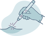
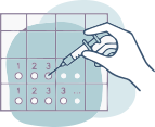
The specialists at Animal Dermatology Clinic are adept at using utilizing surgical procedures to contribute to better treatment outcomes for your pet. And with advanced use of CO2 laser surgery pioneered by Animal Dermatology Clinic dermatologists, we can address challenging dermatological conditions while reducing your pet’s time in surgery and accelerating their recovery.Skin BiopsySkin biopsy, the most common type of surgery performed in pets, is the process of obtaining a small sample of skin or a skin lesion to provide more in-depth information about a particular disease, infection, growth or tumor. This procedure, almost always done outpatient and requiring only local anesthesia, helps more accurately diagnose deep infections, immune mediated diseases, differentiate between benign and malignant growths, and helps meet other diagnostic needs.Biopsies are one of the most informative procedures that can be performed on your pet and, along with expert interpretation of the biopsy sample, may be the difference between identifying the right path toward resolution of a problem and a wrong or incomplete diagnosis.CryosurgeryCryosurgery is a medical procedure for rapidly freezing tissue in order to destroy abnormal or unwanted skin lesions, such as skin tags, warts, cysts, etc. A very cold substance (nitrous oxide) is expertly applied to a pet’s lesion with pinpoint accuracy. The tissue will scab, or 'crust' over within a few days and simply fall off within 14 days of the procedure. Cryosurgery is a safe, quick procedure that does not require sedation and results in little discomfort to your pet.C02 Laser SurgeryAnimal Dermatology Clinic employs CO2 laser surgery to perform more advanced and efficient surgical procedures, often with much less pain on recovery. CO2 lasers are often used in areas where traditional surgeries are not possible, such as within the earl canal, and can also be helpful in areas with good blood supply and where excess bleeding might occur, like the nose. This tool allows our dermatologists to make precise incisions with more limited bleeding than traditional surgery.Since dermatologists often see patients with multiple lesions, CO2 laser surgery is ideally suited for our field. While traditional surgery for multiple lesions can take hours of cutting and suturing each individual site, with a CO2 laser, the procedure can often be done in half the time and the healing is rapid.Specialized surgical procedures performed using a CO2 laser include removal of recurrent draining sores between the toes, ablation of skin cancer such as squamous cell carcinoma in cats, removal of growths or lesions on the pet’s nose and removal of some masses within the ear canal.Find Out if Surgery is an Option for Your Pet’s ConditionWhile many pet owners consider surgery a last resort for their pets, often early surgical intervention may prevent further progression of disease, provide your pet relief and improve their quality of life.The dermatologists and care team at Animal Dermatology Clinic are always happy to discuss whether a specific surgical procedure is the right choice for your pet.CryosurgeryCryosurgery+21 01 Sebaceous adenomas2 02 Sebaceous adenomas3 03 Sebaceous adenomas 10 days post sx

image

Dermatopathology (Skin Biopsy)

For some skin diseases a biopsy can be the only way to reach a definitive diagnosis.Animal Dermatopathology Services (ADS) provides specialized services for expert interpretation of skin biopsies, second opinions on previously collected biopsies and clinicopathologic correlates, and treatment recommendations. Our clinical team, consisting of a board certified pathologist and a board certified dermatologist with extensive and comprehensive experience in the diagnosis and treatment of skin diseases, can help augment your ability to diagnose and treat challenging dermatological cases.ADS uses Pathwood Veterinary Laboratories and Biosphere Lab to process sample submissions. Biopsy samples should be submitted to:West Coast:Pathwood Veterinary Laboratories1415 NE 123rd St.Seattle, WA 98125East Coast: Biosphere Lab (Attn: Dr. Biswell)11700 Commonwealth Dr. Suite 200 Louisville, KY 40299To submit a sample for evaluation:IF SUBMITTING IN WINTER/ FREEZING TEMPS There can be significant artifact if samples are frozen during transit (making histologic interpretation difficult or impossible)For more questions concerning Animal Dermatopathology Services, please contact us at .WHEN ARE BIOPSIES INDICATED?There are several dermatological presentations and considerations that warrant biopsy sampling. Some of the most common include:GENERAL CONSIDERATIONS PRIOR TO BIOPSYBIOPSY PROCEDUREThe biopsy procedure requires a minor surgery with sutures typically used to close the site. Depending on the body location to be sampled and the temperament of the pet, mild sedation or general anesthesia may be needed. Hair overlying the lesion may be gently clipped, but the clipper blades should not touch the skin or crusts. Nodular lesions may be lightly prepared, but do not scrub or prepare crusted, papular, or pustular lesions, as important surface pathology may be damaged.Inject 0.5 to 1 ml of 1% to 2% lidocaine subcutaneously under the lesion using a 25-gage needle. A small amount of 8.4% sodium bicarbonate (0.1 ml/1 ml of lidocaine) can be added to the lidocaine to alleviate any stinging sensation. Make sure you administer the lidocaine subcutaneously; if administered intradermally it can create an artifactual hemorrhage. Allow approximately 5 minutes for the lidocaine to reach full effect before beginning the biopsy.As a rule, do not exceed 1 ml of lidocaine per 5 kg of body weight, and in small dogs and cats care should be used to avoid more than 1 ml on the head and cervical areas. When higher volumes of lidocaine are used in these locations in smaller patients there is a higher incidence of lidocaine reactions. These reactions can create muscle or facial twitching, tremors or seizures. More severe signs with overdosing can include bradycardia, hypotension, and cardiac arrest. Milder reactions are usually self-limiting.For most cases, 6 or 8 mm biopsy punches are preferred but 3-5 mm biopsy punches may be necessary for difficult areas such as near the eye, on the pinnae, and on the nasal planum or footpads of smaller patients. Do not reuse punches as they become dull and tend to shear and distort tissue, creating artifacts. Use of name brand punches (Baker, Miltex or Acuderm) is recommended. Excisional biopsies with a scalpel may be indicated for larger or nodular lesions, or when neoplasia is suspected and for diseases of the subcutaneous fat.Place the punch over the lesion to be sampled and apply even pressure until the punch is fully through the dermis into the subcuticular tissue. Rotating the punch in one direction, as opposed to twisting the punch back and forth, is generally recommended to avoid shearing artifacts. Care should be taken to avoid underlying known vascular structures. As a rule, when using punch biopsy sampling, you want the center of lesion to be your primary focus.Try to avoid large areas of normal skin with the biopsy sample or the lesion may be missed when the biopsy sample is processed and cut, resulting in only normal skin being placed on the slide. If normal skin is wanted for comparison, take a sample from an adjacent area, or use large biopsy punches 8 or 12mm or excisional biopsy samples. The only time a lesion should be biopsied on the margin is in the case of an ulcerative skin disease. If a large lesion appears to be different in the center from the leading edges, obtain biopsy samples from both areas, or use an excisional technique.Use a thumb forceps to grasp the underlying fat, avoiding iatrogenic trauma to the dermis. Handle the sample carefully to avoid any crushing artifact. Place the sample on a gauze sponge and gently absorb any blood from the outer surface of the sample to avoid excess hemorrhage being reported on biopsy sample. Close the biopsy site with simple interrupted sutures or a cruciate suture pattern.PROCESSING SAMPLESAfter obtaining biopsy samples (but before fixation), place the samples on a gauze sponge to absorb excess hemorrhage, and immerse samples in formalin quickly after removal (< 1 min) to help prevent drying artifact.Samples should then be placed in 10% neutral buffered formalin (minimum 10 parts formalin to 1 part tissue for adequate fixation). See note for shipment in cold weather above.Please remember to give a complete signalment and history, including description and distribution of lesions; other signs; the results of pertinent diagnostic tests; current or past therapy and the response to therapy; and your differential diagnoses. These elements are important to allow the pathologist to formulate an accurate diagnosis.For more questions concerning ADG Dermatopathology Services, please contact us at .REFERENCESYager JA, Wilcock BP. Color Atlas and test of surgical pathology of the dog and cat. Dermatopathology and Skin Tumor. London, 1994, Wolfe Publishing.Linder KE. Skin biopsy site selection in small animal dermatology with introduction to histologic pattern analysis of inflammatory skin lesions. Clin Tech Small Anim Paract 16;207-2013, 2001.Gross TL, Ihrke, PJ, Walder EJ. Skin disease of the dog and cat, 2nd ed. Oxford, UK, 2005. Blackwell Science Ltd.Biopsy samples submitted to ADG are processed by Pathwood Veterinary Laboratories(1415 NE 123rd Street, Seattle, WA 98125) and Biosphere Lab (11700 Commonwealth Dr. Suite 200, Louisville, KY 40299).


image

In-House Diagnostics (Cytology, Skin Scrape, Culture)

The good news is that the problem is often literally on the surface. This allows for direct observation of lesions and for diagnostic tests to be performed in a quick and typically painless way. Veterinary dermatologists and the staff at Animal Dermatology Clinic are trained in evaluating these tests on site, in our laboratory to provide results in minutes instead of days. This allows more rapid and accurate decisions on the right course of treatment instead of waiting for test results that may be sent to outside laboratories.Tests performed during a typical examination may include: impression smear cytology, superficial or deep skin scrapes, trichography, fungal cultures and in house blood tests. Depending on the individual pet’s presentation of symptoms, a skin biopsy and dermatopathology reading may be recommended and scheduled.Diagnostic CytologyDiagnostic cytology, also called impression smear cytology, is a common diagnostic test that is used to determine the presence of bacteria, yeast or other infections, inflammation and neoplastic (cancer) cells, and alert the dermatologist to other potential disease processes. Samples for diagnostic cytology are often collected by cotton swab, direct smearing of a glass microscope slide, use of clear tape to collect samples, or in some cases fine needle aspirate to obtain samples from a mass or lesion beneath the skin.By performing this test in-house, veterinary dermatologists can obtain valuable information about a patient or condition while the pet is in the office. This allows for more accurate diagnosis, better decision making on therapies and better patient outcome in managing problem skin diseases.Skin ScrapeSkin scrapes are another useful tool often performed by veterinary dermatologists. Unlike cytology, skin scrapes are performed by using a dulled scalpel blade to scrape along the surface of the skin to collect material for examination under the microscope. Material is placed onto a microscope slide with a small amount of mineral oil and is examined closely for evidence of parasites. Skin scrapes are used primarily to look for mites including Demodex or the mites that cause Scabies.Demodex mites commonly live within hair follicles of the skin and may cause inflammation, hair loss scaling and secondary infections. Since these mites live below the surface of the skin, a “deep” skin scrape is needed to identify them. Deep skin scrapes are often performed in relatively small areas (approx. 1 cm diameter) and typically leave a small superficial abrasion, indicating that the scrape has been done deep enough to find the mites.Superficial skin scrapes involve a broader sweeping scrape done over a larger surface area. This type of scrape is designed to collect surface crust and skin cells and often leave little external evidence that it was performed. Superficial scrapes are used to look for Sarcoptes and other surface mites that may cause inflammation and in many cases intense itching and scratching. Unlike Demodex mites, parasites found on superficial skin scrapes may be elusive with only a few present in a given area making them difficult to find.Skin scrapes can be a game changer since finding mites can dramatically alter the course of treatment and often leads to much more rapid resolution of skin disease and discomfort.TrichographyTrichography is the microscopic examination of hairs. Normal hairs of animals have well documented appearance and structure. By plucking hairs from an animal with skin or haircoat disease, a veterinary dermatologist can evaluate the hairs and look for evidence of infection, trauma or malformation. This can also identify parasites like Demodex that live within the hair follicle.Dermatophyte TestingDermatophytosis (ringworm) is a fungal infection of the hair, superficial skin, and occasionally nails. It’s not caused by a worm and often lesions are not in rings. Dermatophytosis is instead caused by infection with one of three species of fungal organism: Microsporum canis, Microsporum gypseum, and Trichophyton mentagrophytes.Common sources of infection include infected pets, rodents, contaminated environments, and occasionally the soil. While less common than many other skin diseases, dermatophytosis is a contagious and zoonotic disease that can in some cases be transmitted to humans. This makes it an important disease to assess and diagnose when the index of suspicion is high. Methods for diagnosing dermatophytosis include fungal culture, PCR testing and on occasion skin biopsy. In some cases, fungal spores may be identified on impression smear cytology raising the suspicion of dermatophytosis. Woods lamp examination may also increase suspicion of dermatophytosis.If a veterinary dermatologist feels it is appropriate to test for dermatophytosis, most often he or she will collect hair and skin samples from the area(s) involved for one of these tests. Identification of the specific fungal organism responsible for skin lesions via fungal culture is the most reliable of these methods. Fungal cultures often take 14 to 21 days to complete to allow identifiable fungal organism growth.When treating a pet for dermatophytosis it is imperative to treat the animal beyond clinical cure, when no further lesions can be seen. To confirm that the infection has been completely resolved a fungal culture is typically repeated to confirm successful therapy.In-House Blood/Urine TestingSkin disease in animals can sometimes be an external marker of an internal problem. Examples include disease such as Hypothyroidism, Cushing’s disease and others. For these conditions, information provided by bloodwork or urinalysis can be vital to reaching a diagnosis and directing a proper course of treatment. Animal Dermatology Clinics perform many blood tests in our in-house laboratory, providing more rapid information and diagnosis.Skin Biopsy and DermatopathologyTo perform a biopsy is to surgically remove a portion of diseased skin, tissue or tumor in order to further analyze this tissue and understand the disease process more completely. For some skin diseases, this is the only accurate way to differentiate one disorder from another and can be the difference between accurate diagnosis and the wrong direction.To perform a skin biopsy, most animals are sedated or anesthetized and often a local anesthetic is used to provide further anesthesia and pain control. Many skin biopsies are performed using a “punch” biopsy instrument - a small circular blade that may vary from 4 to 8 mm. Use of a biopsy punch allows a small sample to be obtained from areas of concern and are often sutured with a single stitch.What happens to biopsy skin or tissue after this procedure is performed is of equal importance as the procedure itself. Animal Dermatology Clinic is fortunate to have an in-house dermatopathology service to interpret findings from the biopsy once the tissue has been processed. Dr. Wayne Rosenkrantz, DVM, DACVD, and Dr. Kelly Keating, DVM, DACVP provide dermatopathology interpretation services to both Animal Dermatology Clinic dermatologists, and other specialists and general practitioners. For veterinarians interested in learning more about this service, click here.Diagnostics - Biopsy 1 - Biopsy procedure Image 2 300dpi[1]-1In-House Diagnostics-1-1Diagnostics - Cytology 2 - Bacteria TNTC

spec
Visit

Location & Hours

Address19035 W. Capitol Drive, Suite J-100, Brookfield, WI 53045
Monday2:30 am – 11:30 am
Tuesday2:30 am – 11:30 am
Wednesday2:30 am – 11:30 am
Thursday2:30 am – 11:30 am
ClosedFri, Sat, Sun
Visit their website to contact them
Open in Google Maps

Explore Great Lakes Veterinary Dermatology

prep-surgery.jpeg
For referring veterinarians

Refer a patient to Great Lakes Veterinary Dermatology

Send your case directly to Great Lakes Veterinary Dermatology and stay in the loop on your patient's care.

Refer a patient
ophtho
For specialists

Join the Network

Interested in joining the fastest-growing specialty network? See what AOSN offers.

Learn about joining AOSN