Dermatopathology (Skin Biopsy)
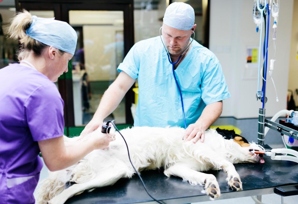
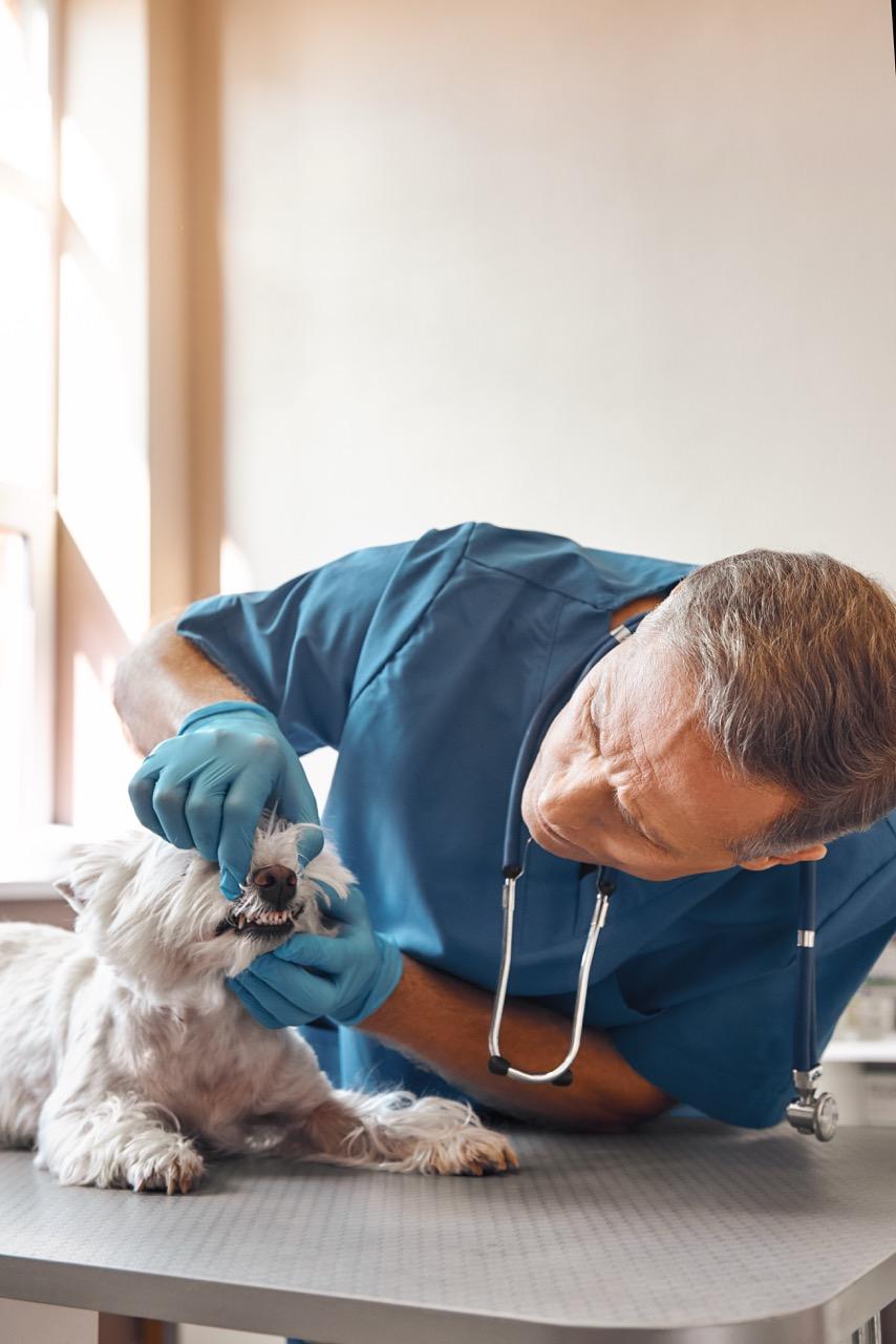
For some skin diseases a biopsy can be the only way to reach a definitive diagnosis.Animal Dermatopathology Services (ADS) provides specialized services for expert interpretation of skin biopsies, second opinions on previously collected biopsies and clinicopathologic correlates, and treatment recommendations. Our clinical team, consisting of a board certified pathologist and a board certified dermatologist with extensive and comprehensive experience in the diagnosis and treatment of skin diseases, can help augment your ability to diagnose and treat challenging dermatological cases.ADS uses Pathwood Veterinary Laboratories and Biosphere Lab to process sample submissions. Biopsy samples should be submitted to:West Coast:Pathwood Veterinary Laboratories1415 NE 123rd St.Seattle, WA 98125East Coast: Biosphere Lab (Attn: Dr. Biswell)11700 Commonwealth Dr. Suite 200 Louisville, KY 40299To submit a sample for evaluation:IF SUBMITTING IN WINTER/ FREEZING TEMPS There can be significant artifact if samples are frozen during transit (making histologic interpretation difficult or impossible)For more questions concerning Animal Dermatopathology Services, please contact us at .WHEN ARE BIOPSIES INDICATED?There are several dermatological presentations and considerations that warrant biopsy sampling. Some of the most common include:GENERAL CONSIDERATIONS PRIOR TO BIOPSYBIOPSY PROCEDUREThe biopsy procedure requires a minor surgery with sutures typically used to close the site. Depending on the body location to be sampled and the temperament of the pet, mild sedation or general anesthesia may be needed. Hair overlying the lesion may be gently clipped, but the clipper blades should not touch the skin or crusts. Nodular lesions may be lightly prepared, but do not scrub or prepare crusted, papular, or pustular lesions, as important surface pathology may be damaged.Inject 0.5 to 1 ml of 1% to 2% lidocaine subcutaneously under the lesion using a 25-gage needle. A small amount of 8.4% sodium bicarbonate (0.1 ml/1 ml of lidocaine) can be added to the lidocaine to alleviate any stinging sensation. Make sure you administer the lidocaine subcutaneously; if administered intradermally it can create an artifactual hemorrhage. Allow approximately 5 minutes for the lidocaine to reach full effect before beginning the biopsy.As a rule, do not exceed 1 ml of lidocaine per 5 kg of body weight, and in small dogs and cats care should be used to avoid more than 1 ml on the head and cervical areas. When higher volumes of lidocaine are used in these locations in smaller patients there is a higher incidence of lidocaine reactions. These reactions can create muscle or facial twitching, tremors or seizures. More severe signs with overdosing can include bradycardia, hypotension, and cardiac arrest. Milder reactions are usually self-limiting.For most cases, 6 or 8 mm biopsy punches are preferred but 3-5 mm biopsy punches may be necessary for difficult areas such as near the eye, on the pinnae, and on the nasal planum or footpads of smaller patients. Do not reuse punches as they become dull and tend to shear and distort tissue, creating artifacts. Use of name brand punches (Baker, Miltex or Acuderm) is recommended. Excisional biopsies with a scalpel may be indicated for larger or nodular lesions, or when neoplasia is suspected and for diseases of the subcutaneous fat.Place the punch over the lesion to be sampled and apply even pressure until the punch is fully through the dermis into the subcuticular tissue. Rotating the punch in one direction, as opposed to twisting the punch back and forth, is generally recommended to avoid shearing artifacts. Care should be taken to avoid underlying known vascular structures. As a rule, when using punch biopsy sampling, you want the center of lesion to be your primary focus.Try to avoid large areas of normal skin with the biopsy sample or the lesion may be missed when the biopsy sample is processed and cut, resulting in only normal skin being placed on the slide. If normal skin is wanted for comparison, take a sample from an adjacent area, or use large biopsy punches 8 or 12mm or excisional biopsy samples. The only time a lesion should be biopsied on the margin is in the case of an ulcerative skin disease. If a large lesion appears to be different in the center from the leading edges, obtain biopsy samples from both areas, or use an excisional technique.Use a thumb forceps to grasp the underlying fat, avoiding iatrogenic trauma to the dermis. Handle the sample carefully to avoid any crushing artifact. Place the sample on a gauze sponge and gently absorb any blood from the outer surface of the sample to avoid excess hemorrhage being reported on biopsy sample. Close the biopsy site with simple interrupted sutures or a cruciate suture pattern.PROCESSING SAMPLESAfter obtaining biopsy samples (but before fixation), place the samples on a gauze sponge to absorb excess hemorrhage, and immerse samples in formalin quickly after removal (< 1 min) to help prevent drying artifact.Samples should then be placed in 10% neutral buffered formalin (minimum 10 parts formalin to 1 part tissue for adequate fixation). See note for shipment in cold weather above.Please remember to give a complete signalment and history, including description and distribution of lesions; other signs; the results of pertinent diagnostic tests; current or past therapy and the response to therapy; and your differential diagnoses. These elements are important to allow the pathologist to formulate an accurate diagnosis.For more questions concerning ADG Dermatopathology Services, please contact us at .REFERENCESYager JA, Wilcock BP. Color Atlas and test of surgical pathology of the dog and cat. Dermatopathology and Skin Tumor. London, 1994, Wolfe Publishing.Linder KE. Skin biopsy site selection in small animal dermatology with introduction to histologic pattern analysis of inflammatory skin lesions. Clin Tech Small Anim Paract 16;207-2013, 2001.Gross TL, Ihrke, PJ, Walder EJ. Skin disease of the dog and cat, 2nd ed. Oxford, UK, 2005. Blackwell Science Ltd.Biopsy samples submitted to ADG are processed by Pathwood Veterinary Laboratories(1415 NE 123rd Street, Seattle, WA 98125) and Biosphere Lab (11700 Commonwealth Dr. Suite 200, Louisville, KY 40299).